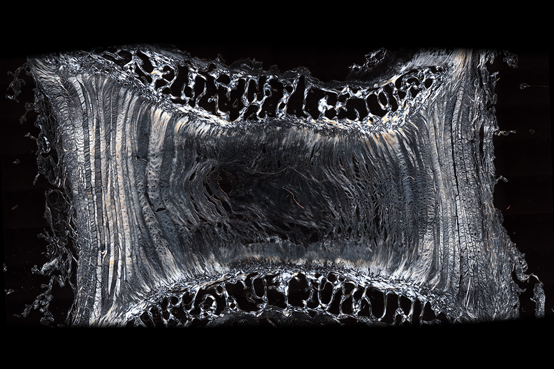
A major part of this work is in the use of multi-axis spine testing. I have led in the development of a custom six-axis test system that can replicate the complex loads and movements of the spine, rather than test regimes that use simple waveforms in one axis at a time. Following this, a biochamber was integrated onto the six-axis test system so that we can apply complex physiological activity profiles to whole organ intervertebral disc cultures. This allows us to complete research into interverebral disc mechanobiology in ways not previously possible, and provides a unique system for the evaluation of novel treatments and therapies for the spine.

Another area of research is investigating degenerative mechanisms in the intervertebral disc, and developing ways to replicate the characteristics of degeneration in the in vitro environment. This work runs alongside our mechanobiology research using the six-axis bioreactor, so that we can understand more about how degeneration occurs and progresses, and provide better ways to evaluate strategies to prevent, manage, and treat it.

| 
| 
|
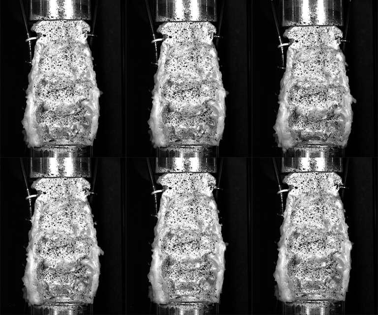
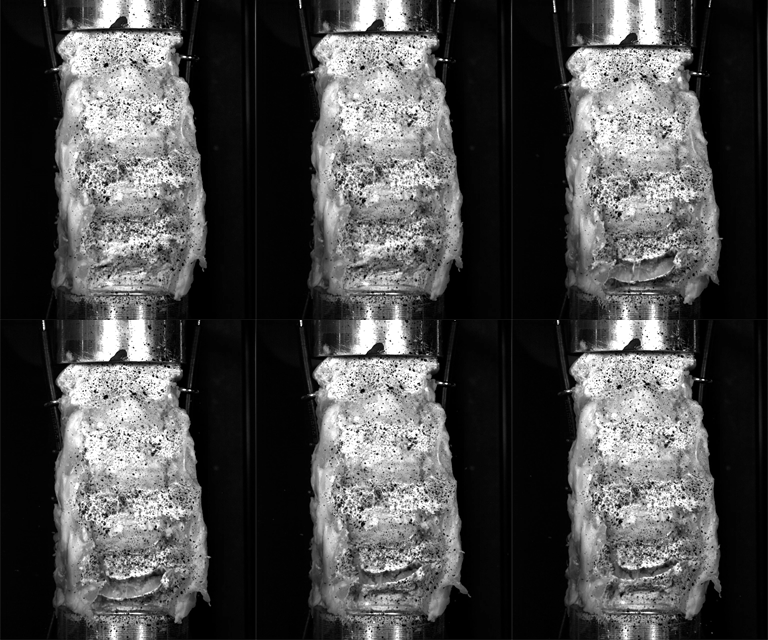
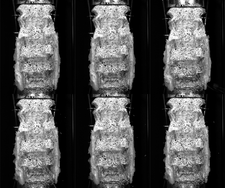
Understanding the mechanisms of injury is really important if we want to develop ways to minimise risk, whether that is through preventative measures such a rule/law changes, education, or the development of safety devices. I have been involved in research relating to cervical spine injuries during to car collisions, and in Rugby Union. As with much of my research, a critical part of the work was in developing the test systems to replicate the movement or loading on the spine in a realistic manner, so that the data collected was relevant to the problem. The above tests replicated the loading during Rugby Union scrummaging that if directed onto the cervical spine could lead to catastrophic injury. Because the impacts occur over only a few milliseconds, we used high-speed (4000 frames per second) 3D digital image correlation to measure the surface bone strain and vertebral motion during the impacts. This data helped to understand the way that the spine behaved under these high impact loads, and was also used to validate musculoskeletal models, which could then be used for further investigations of spinal loading, and injury risk.